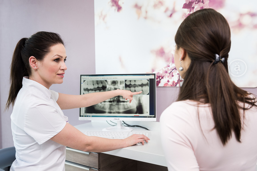
Dental X-Ray
X-ray is an auxiliary diagnostic method that uses X-radiation and its varying ability to penetrate soft and hard tissues. This examination method allows us to see places that are normally invisible. Different types of imaging show different types of hard layers (bones, teeth). We use digital radiovisiography in our practice. X-radiation is not captured on a film, but on a computer sensor that displays the image directly on the computer screen. This technology reduces radiation doses by a factor of ten compared to conventional imaging. We can edit the image, zoom in on details and archive the image on a CD.
Intraoral X-ray
A small intraoral image inside the mouth, focused on one or two teeth and their vicinity.
OPG
Orthopantomogram is a large image showing the lower and upper jaw and nasal cavities and the temporomandibular joint (the jaw-temple joint).
TELE X-RAY
It is an image used in orthodontic analysis. It shows the entire skull in the side projection, which is important for accurate determination of treatment.
BITEWING X-RAY
It is an intraoral image of three to four upper or lower teeth focused on decay diagnostics in interdental spaces.


